
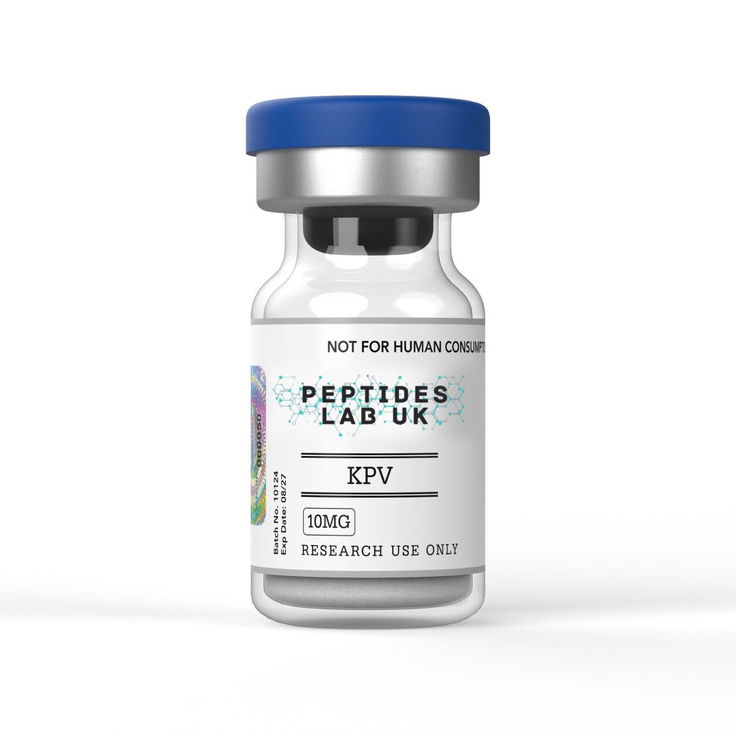
KPV peptide is the C-terminal tripeptide fragment of alpha-melanocyte-stimulating hormone (α-MSH), consisting of the amino-acid sequence Lys-Pro-Val (KPV; α-MSH residues 11–13). Unlike the complete 13-amino-acid melanocortin peptide, KPV is an exceptionally small molecular system whose biological activity has been investigated primarily in cellular, biochemical and preclinical models. Its importance in research derives from the observation that selected biological effects associated with α-MSH can be reproduced by its C-terminal KPV sequence without reproducing the complete peptide architecture. Experimental work has investigated KPV in relation to epithelial peptide transport, NF-κB signalling, MAP-kinase pathways, cytokine expression and microbial interactions. These findings provide a mechanistic framework for studying peptide-mediated regulation of inflammatory signalling, but they do not establish clinical efficacy, safety or therapeutic use.
What Is KPV Peptide?
KPV is a linear tripeptide composed of Lysine-Proline-Valine, conventionally abbreviated K-P-V. It corresponds to residues 11–13 at the C-terminal end of α-MSH, whose complete sequence is SYSMEHFRWGKPV. α-MSH itself is derived from the pro-opiomelanocortin (POMC) precursor and belongs to the melanocortin peptide family. The final three residues therefore represent a highly compact fragment of a much larger signalling molecule.
The structural significance of KPV is greater than its length might suggest. A tripeptide contains only three peptide bonds and a relatively small number of functional groups, yet the sequence combines a basic lysine residue with the conformationally influential cyclic amino acid proline and the hydrophobic branched-chain amino acid valine. This produces a molecular structure substantially simpler than α-MSH and allows researchers to investigate whether particular biological effects depend on the complete melanocortin sequence or can arise from a smaller terminal motif.
A particularly important distinction concerns pigmentation. The canonical melanocortin receptor mechanism of α-MSH involves receptor binding and downstream cyclic-AMP signalling, especially through melanocortin receptors expressed by melanocytes. Experimental research has found that KPV behaves differently from the receptor-active core of α-MSH. In keratinocyte studies, KPV did not reproduce the expected cyclic-AMP response associated with conventional melanocortin receptor signalling. Other work has likewise indicated that the anti-inflammatory behaviour of KPV can occur independently of classical melanocortin receptor activation.
This distinction is central to KPV research because it separates sequence-derived intracellular activity from the canonical endocrine-style receptor activity of intact α-MSH. The literature consequently treats KPV as a useful molecular probe for investigating melanocortin-derived signalling without assuming that every biological property of α-MSH is retained.
Another important characteristic is peptide transport. KPV is sufficiently small to interact with peptide transport systems that normally recognise di- and tripeptides. Research using intestinal epithelial models identified peptide transporter 1 (PepT1) as an important route for KPV uptake. In Caco-2-BBE and HT29-Cl.19A cells, radiolabelled KPV uptake experiments supported active PepT1-mediated transport, with kinetic experiments reporting a Km of approximately 160 μM under the experimental conditions used.
This transport mechanism has major implications for laboratory experiments. KPV cannot simply be treated as a generic extracellular signalling molecule. Its cellular effects depend, at least in some models, on the ability of the peptide to cross the plasma membrane through an appropriate transporter and reach intracellular compartments.
Claims about mucosal penetration therefore need to be interpreted carefully. Published work demonstrates cellular uptake and transport in experimental epithelial systems, rather than establishing a human absorption profile. A fluorescent KPV-containing probe has also been used experimentally to visualise PepT1-associated accumulation in intestinal epithelial models, providing further evidence that the KPV motif can function as a molecular recognition element for peptide transport.
From a structural perspective, KPV is therefore interesting precisely because it is small: its low molecular complexity facilitates controlled structure–activity investigations, peptide-transport experiments and analytical characterisation while allowing researchers to distinguish sequence-specific effects from the broader pharmacology of intact α-MSH.
Mechanism of Action
The most extensively investigated KPV mechanism involves intracellular modulation of inflammatory signalling rather than conventional melanocortin-receptor activation. The pathway can be conceptualised as a sequence of events beginning with PepT1 recognition, followed by cellular uptake and subsequent interference with transcription-factor trafficking.
PepT1 is a proton-coupled oligopeptide transporter capable of transporting di- and tripeptides. In the intestine, it is normally associated with nutrient peptide absorption, while experimental inflammatory conditions can alter its expression in intestinal epithelial tissues. The seminal KPV transport study demonstrated that KPV competes with established PepT1 substrates and that radiolabelled KPV is transported into epithelial and immune-cell models. Blocking the transporter with competing substrates attenuated KPV-associated signalling effects, supporting a transporter-dependent mechanism.
Once intracellular, KPV has been associated with suppression of NF-κB activation. NF-κB is a transcription-factor system that controls the expression of numerous inflammatory and immune-response genes. In a canonical pathway, inflammatory stimuli activate the IKK complex, leading to phosphorylation and subsequent degradation of IκB proteins. Liberation of NF-κB, particularly the p65/RelA-containing complex, permits nuclear translocation and transcriptional activation.
KPV appears to interfere with this process at more than one level. In intestinal epithelial-cell experiments, KPV reduced stimulus-associated IκB-α degradation and altered the duration of NF-κB activation. NF-κB reporter assays, immunoblotting and electrophoretic mobility-shift experiments were used to demonstrate the effect. The same research linked KPV activity to PepT1 expression, because transporter competition or absence of PepT1 substantially reduced the observed signalling response.
A separate mechanistic investigation in human bronchial epithelial cells provided additional insight into intracellular trafficking. The researchers reported that KPV accumulates predominantly in the nucleus and interferes with the interaction between NF-κB p65/RelA and importin-α3. This reduces p65 nuclear translocation and changes the kinetics of NF-κB activation. The study also reported increased cellular abundance of IκB-α following KPV exposure.
It is important to distinguish nuclear translocation from nucleolar translocation. The available mechanistic evidence supports KPV accumulation in the nucleus and interference with nuclear trafficking of NF-κB; it does not establish a specific KPV-mediated nucleolar-translocation mechanism. For scientifically accurate reporting, the process should therefore be described as nuclear rather than nucleolar trafficking.
Downstream consequences have been investigated through cytokine and chemokine measurements. In intestinal epithelial models, KPV exposure reduced signalling associated with pro-inflammatory cytokine stimulation and decreased IL-8 secretion. Published animal experiments additionally measured changes in inflammatory cytokine mRNA. The literature therefore supports a model in which PepT1-mediated uptake is upstream of intracellular signalling changes, with NF-κB and MAPK pathways representing important downstream analytical endpoints.
The frequently discussed TNF-α/IL-6 axis should also be interpreted cautiously. TNF-α has been used experimentally as an inflammatory stimulus in KPV mechanistic studies, particularly in epithelial models. However, cytokine responses are model-dependent, and not every KPV experiment measures both TNF-α and IL-6 directly. A scientifically defensible description is therefore that KPV research has examined modulation of inflammatory cytokine programmes, including TNF-associated and interleukin-associated signalling, rather than claiming a universal cytokine-suppression mechanism.
KPV research also intersects with antimicrobial biology. α-MSH and its C-terminal KPV fragment have been investigated against organisms including Staphylococcus aureus and Candida albicans. One study reported that both α-MSH and KPV influenced microbial viability and that cAMP-related mechanisms could contribute to the observed activity in yeast.
This antimicrobial literature should not be conflated with the PepT1/NF-κB mechanism. The two areas represent different experimental systems: one concerns peptide uptake and intracellular inflammatory signalling in mammalian cells, while the other concerns direct peptide–microorganism interactions. The antimicrobial behaviour of α-MSH-related peptides has also been associated with membrane interactions, cAMP perturbation and sequence-dependent structural effects.
Taken together, the mechanistic picture is therefore multi-layered: PepT1-mediated uptake → intracellular accumulation → altered NF-κB trafficking/activation and MAPK signalling → measurable changes in inflammatory gene and cytokine readouts, alongside separate experimental evidence for direct antimicrobial interactions. None of these mechanisms should be interpreted as evidence of clinical efficacy.
What the Research Shows
The published literature provides several useful experimental models for understanding KPV.
1. PepT1-mediated intestinal epithelial transport
Dalmasso and colleagues investigated KPV using Caco-2-BBE and HT29-Cl.19A intestinal epithelial cells together with Jurkat T-cell models. The study combined NF-κB reporter assays, Western blotting, RT-PCR, ELISA and radiolabelled peptide uptake experiments. KPV was transported by PepT1, and transporter competition reduced its signalling effects. The investigators also examined DSS- and TNBS-induced murine colitis as preclinical models. Importantly, the dosing and administration reported in that work belong exclusively to the experimental animal model and should not be extrapolated into human-use protocols.
PubMed — PepT1-mediated tripeptide KPV uptake reduces intestinal inflammation
2. Murine intestinal inflammation models
A separate investigation examined KPV in two experimental models of intestinal inflammation: DSS-induced colitis and CD45RB^hi T-cell-transfer colitis. The researchers also investigated animals with non-functional MC1 receptors to help determine whether KPV required conventional melanocortin receptor signalling. The study reported experimental anti-inflammatory effects while providing evidence that KPV could operate independently of functional MC1R signalling.
This distinction is scientifically valuable because it helps separate KPV from the receptor-dependent pharmacology of intact α-MSH. It also illustrates why animal observations should remain classified as preclinical findings rather than human therapeutic evidence.
3. Keratinocyte signalling
Research in human keratinocyte systems examined α-MSH, KPV and related peptides. The investigators found that KPV did not produce the expected cyclic-AMP response in HaCaT or normal keratinocytes under the tested conditions, although intracellular calcium responses were observed in particular experimental settings. These findings support the view that KPV cannot simply be assumed to reproduce the full MC1R/cAMP signalling profile of α-MSH.
PubMed — α-MSH, KPV and signalling in human keratinocytes
4. Nuclear NF-κB trafficking
The bronchial epithelial-cell study provides perhaps the clearest mechanistic evidence for intracellular KPV action beyond the intestinal system. KPV was observed intracellularly and was associated with inhibition of p65/RelA nuclear translocation. The proposed mechanism involved competition around importin-mediated nuclear transport and changes in IκB-α abundance. These experiments reinforce the concept that KPV can influence inflammatory signalling through intracellular protein-trafficking mechanisms rather than simply acting as a conventional extracellular receptor agonist.
5. Antimicrobial interactions
Early research investigated α-MSH peptides, including KPV, against S. aureus and C. albicans. The work demonstrated antimicrobial influences at experimental concentrations and proposed cAMP-related mechanisms in yeast. Later research into α-MSH-derived analogues expanded the investigation into peptide structure, charge and microbial membrane interactions. These studies are useful for structure–activity research but should not be interpreted as evidence that KPV is an antimicrobial treatment.
PMC — Antimicrobial properties of α-MSH and related synthetic melanocortins
Collectively, the literature establishes a coherent preclinical research theme: KPV is a small α-MSH-derived peptide that can participate in PepT1-dependent intracellular transport, influence inflammatory signalling pathways and exhibit experimentally measurable interactions with microorganisms. However, these findings remain dependent on experimental system, cell type, peptide preparation, concentration and assay design.
Research Applications
KPV is particularly useful as a mechanistic laboratory tool because its short sequence permits researchers to isolate questions that would be difficult to answer using the complete α-MSH molecule. Experimental applications can be divided into several categories.
The first is intestinal epithelial-cell research. Caco-2-BBE and HT29-derived systems provide established models for examining epithelial transport, barrier-associated signalling and inflammatory responses. KPV can be incorporated into experiments designed to compare transporter-positive and transporter-deficient cells. Appropriate controls may include established PepT1 substrates, transporter competition experiments and vehicle-treated controls.
A second application is NF-κB pathway analysis. Reporter constructs containing NF-κB-responsive elements can provide quantitative measurements of transcription-factor activation following inflammatory stimulation. Western blotting can complement reporter assays by measuring IκB-α abundance or phosphorylation-related endpoints, while immunofluorescence or confocal microscopy can investigate p65 localisation. These complementary methods help distinguish transcriptional changes from alterations in intracellular trafficking.
A third research area is cytokine profiling. Researchers can measure transcript and protein endpoints using RT-qPCR, ELISA or multiplex immunoassays. Depending on the model, relevant markers may include IL-1β, IL-6, IL-8, TNF-α and other inflammatory mediators. Experimental interpretation should always distinguish between direct measurement and inferred pathway activity; a reduction in one cytokine does not automatically establish global suppression of inflammation.
KPV is also relevant to mucosal transport assays. Because PepT1 is a transporter for short peptides, researchers can investigate uptake kinetics, competitive inhibition and transporter expression. Radiolabelled peptide approaches have historically been used, while fluorescent conjugates can provide imaging-based alternatives. Importantly, modifying KPV with a fluorophore can change its molecular properties, so labelled constructs should not automatically be assumed to behave identically to native KPV.
When selecting a certified KPV research peptide for intestinal mucosal or cellular transport models, researchers should prioritise documented identity, chromatographic purity, mass confirmation and batch traceability rather than relying solely on nominal peptide content.
Another application is comparative structure–activity research. Researchers can compare KPV with α-MSH, related tripeptide analogues or chemically modified derivatives. Such comparisons can help determine whether a biological endpoint depends on peptide length, terminal chemistry, stereochemistry, charge distribution or receptor interaction. For example, literature examining modified KPV structures demonstrates that even relatively small chemical changes can alter proteolytic stability and antimicrobial behaviour.
Cutaneous epithelial models offer another research context. Keratinocytes can be used to study intracellular signalling, cytokine responses and melanocortin-related pathways. Here, KPV is especially useful as a comparator against intact α-MSH because experiments can separate receptor-mediated melanocortin activity from responses associated with the smaller C-terminal sequence.
Finally, KPV can function as a negative or mechanistic control in transporter experiments. If a response disappears when PepT1 is competitively inhibited or genetically reduced, the resulting comparison can strengthen the interpretation that the experimental effect depends on peptide transport. This type of experimental design is considerably more informative than simply comparing treated and untreated wells.
The overarching research value of KPV therefore lies not in a presumed clinical application but in its utility as a defined molecular probe for studying peptide transport, intracellular trafficking, NF-κB regulation, cytokine signalling and sequence-dependent biological activity.
Purity, Storage and Handling
Analytical quality is particularly important for short peptides because small quantities of truncated sequences, oxidation products, deletion sequences or synthesis-related impurities can influence experimental reproducibility. A nominally labelled KPV sample should therefore be evaluated using orthogonal analytical techniques wherever possible.
For laboratory research, a HPLC purity specification of ≥98% is a commonly appropriate quality benchmark for high-purity peptide material. HPLC, particularly reverse-phase HPLC, can separate the principal peptide from synthesis-related impurities and provide a chromatographic purity estimate. However, chromatographic purity alone does not prove molecular identity. Two compounds with similar retention characteristics can potentially contribute to the same chromatographic region.
This is why mass spectrometry should complement HPLC. LC-MS or MALDI-TOF MS can provide molecular-mass confirmation and help establish that the dominant chromatographic peak corresponds to the expected peptide. For KPV, the theoretical molecular mass depends on the precise chemical form being tested. Native free-acid KPV, acetylated KPV and amidated derivatives are chemically distinct materials and should never be treated as interchangeable simply because they share the KPV sequence.
When evaluating high-purity KPV peptide for laboratory assays, researchers should verify that the batch documentation includes an identity assessment, chromatographic purity result, mass-spectrometric confirmation where available, lot number and relevant storage information.
For an unmodified research peptide supplied in lyophilised form, storage at approximately −20°C is a conventional long-term laboratory storage condition. The material should be protected from unnecessary moisture, repeated temperature cycling and prolonged exposure to ambient laboratory conditions. Small aliquots can be useful where repeated opening of a primary container would otherwise expose the material to humidity and temperature fluctuations.
Reconstitution is a laboratory procedure and should be performed according to the validated specifications of the particular material and experimental protocol. Where aqueous reconstitution is appropriate, researchers may use a compatible sterile buffer or other validated laboratory solvent system. The choice of solvent can influence peptide solubility, aggregation, pH-dependent charge state and downstream assay performance. Consequently, solvent selection should be established experimentally rather than assumed to be universal.
Researchers should also record the exact concentration prepared, solvent composition, preparation date and lot number. This information is especially valuable when comparing experiments performed on different days or using different peptide batches.
Freeze–thaw exposure should be minimised because repeated temperature cycling can increase the opportunity for degradation or adsorption-related losses. Aliquoting prepared laboratory solutions can therefore improve consistency where the experimental design requires repeated measurements.
Analytical verification should ideally be performed on the material as supplied and, where experimental integrity requires it, on prepared solutions. HPLC can provide evidence of chromatographic integrity, while MS can identify unexpected molecular-weight changes. For mechanistic work, additional techniques such as LC-MS/MS can provide higher-resolution confirmation of degradation products.
Importantly, ≥98% purity does not mean 98% biological activity, nor does it establish sterility, endotoxin status or suitability for any biological application beyond the validated laboratory system. Each of these characteristics requires separate analytical controls. Endotoxin testing can be relevant to cell-based inflammatory assays because contaminating bacterial lipopolysaccharide can itself activate innate immune pathways and confound NF-κB or cytokine measurements.
The most rigorous KPV experiments therefore treat quality control as an integrated process: identity confirmation + chromatographic purity + molecular-mass confirmation + appropriate storage + controlled preparation + experimental controls. This approach reduces the likelihood that an apparent biological effect is actually attributable to an impurity, degradation product, solvent effect or handling artefact.
Frequently Asked QuestionsWhat is the amino-acid sequence of KPV peptide?
KPV is a three-amino-acid peptide consisting of lysine (K), proline (P) and valine (V). It corresponds to residues 11–13 at the C-terminal end of α-MSH. Its small size makes it useful for structure–activity, transporter and intracellular-signalling research. KPV should not automatically be considered pharmacologically equivalent to full-length α-MSH because the larger peptide contains additional residues required for canonical melanocortin-receptor interactions.
How does PepT1 transport KPV?
PepT1 is a proton-coupled transporter capable of recognising short peptides. Experimental work using intestinal epithelial cells demonstrated that KPV can be transported through PepT1 and that transporter competition can reduce KPV-associated signalling effects. Radiolabelled uptake experiments were used to characterise this process. The exact transport behaviour remains dependent on cell type, transporter expression, substrate concentration and experimental conditions.
Is KPV structurally stable?
KPV is a very short peptide, but small size does not mean complete resistance to degradation. Peptides can undergo hydrolysis, proteolytic cleavage, oxidation or other chemical changes depending on their sequence, formulation and environmental conditions. Research involving modified KPV analogues demonstrates that relatively small structural changes can substantially alter proteolytic stability. Therefore, stability should be experimentally verified rather than inferred solely from the tripeptide sequence.
How should KPV be handled in laboratory research?
Research-grade KPV should be handled according to the supplier’s certificate of analysis and the laboratory’s validated peptide procedures. Lyophilised material is commonly maintained under frozen conditions such as −20°C, protected from moisture and unnecessary temperature cycling. Where reconstitution is required for an approved in-vitro assay, researchers should use a validated compatible sterile buffer or solvent and document preparation conditions, concentration and lot information.
Conclusion
KPV is a highly compact α-MSH-derived tripeptide whose scientific value comes from the ability to investigate peptide transport and intracellular signalling independently from many properties of the complete melanocortin molecule. Published preclinical research has linked KPV to PepT1-mediated cellular uptake, NF-κB and MAPK signalling, nuclear trafficking and experimentally observed antimicrobial interactions. The evidence remains model-specific and preclinical, making rigorous analytical verification and appropriate experimental controls essential.
Research-use disclaimer: KPV is discussed here strictly as a laboratory research compound. The information above concerns in-vitro, biochemical and preclinical research only and does not provide human dosing, administration or treatment guidance. KPV is not presented as suitable for human or veterinary use.